
Figure 4.
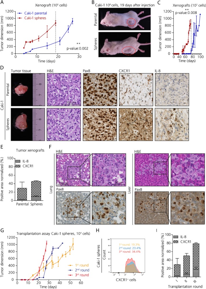
IL‐8/CXCR1 signaling contributes to tumor development and metastasis formation in vivo. (A) Tumor growth evaluation in NSG xenografts subcutaneously injected with 106 cells derived from Caki‐1 spheres (in red) and parental cells (in blue) (unpaired t‐test, n = 3). (B) Representative pictures of immune compromised mice at 19 days post‐injection with either parental or sphere cells. (C) Tumor growth evaluation in NSG xenografts subcutaneously injected with 102 cells derived from Caki‐1 spheres (in red) and parental cells (in blue) (unpaired t‐test, n = 3). (D) Sections of xenografted tumors derived from Caki‐1 showing the histological subtype by H&E, and the kidney nature PAX8 positivity, and differential CXCR1 and IL‐8 expression in the tumors derived from spheres compared to those derived from parental cells. Scale bar: 100 μm. (E) Quantification of immunohistochemical stains for IL‐8 and CXCR1 in tumor xenografts derived from Caki‐1. (F) Micrometastases in lungs and liver of xenografts derived from the injection with 104 Caki‐1 sphere cells. Scale bar: 100 μm, 50 μm. (G) Transplantation assay (n = 3). (H) Analysis using FACS of the CXCR1+ population in xenografted tumors following retransplantation of xenografted tumors. (I) Quantification of immunohistochemical staining for CXCR1 in tumor xenografts derived from Caki‐1 upon retransplantation of xenografted tumors (n = 2).