
Figure 4.
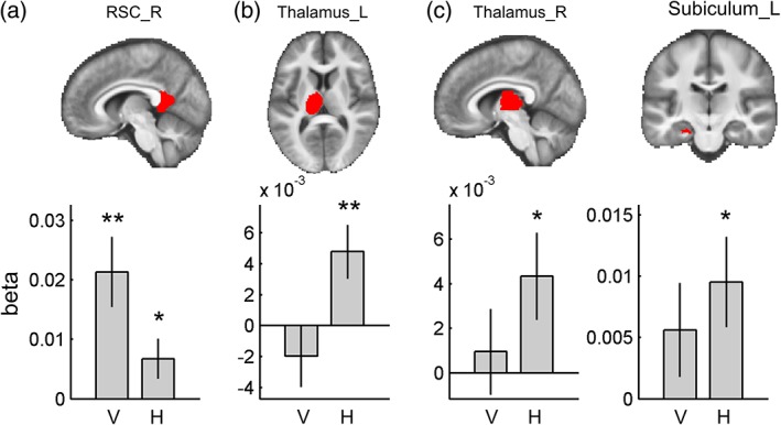
Multivoxel pattern analysis in the ROIs. Each ROI is overlaid on the group‐averaged structural MR image on the top row. (a) Right RSC showed both vertical and horizontal direction encoding, but it was more sensitive to vertical direction. Bilateral thalamus (b, c) and left subiculum (d) showed only horizontal direction encoding. V, vertical; H, horizontal; R, right; and L, left. Error bars are SEM. ** p < .01, * p < .05 [Color figure can be viewed at wileyonlinelibrary.com]