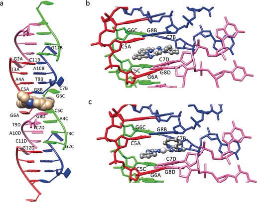
Figure 2.

Crystal structure of the adduct 1–CGTACG. a) B‐Type double‐helix units arranged in a column in which each B‐type double‐helix unit is formed by two contiguous ds‐CGTACG moieties. Complex 1 is shown in CPK style. b, c) Binding site of the platinum complex 1 in the 1‐F and 1‐G conformation, respectively (see text). The four colours red, blue, green, and pink of the DNA strands correspond to the four crystallographically independent DNA oligomers (denoted A–D).
