
Figure 3.
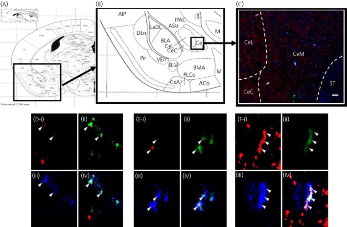
Some glucagon‐like peptide‐1 (GLP‐1) fibres form synapses with interleukin (IL)‐6‐immunoreactive cells in the central amygdala (CeA). GLP‐1 (green), synaptophysin (red) and IL‐6 (blue) was found to co‐localise in some structures of the CeA. Anatomical overview images (A, B) showing the region from which the magnifications were taken. Representative image (C) of the region showing partial co‐localisation of GLP‐1, IL‐6 and synaptophysin. Triple‐channel confocal micrographs (D‐F) showing micrographs of single cells with synaptophysin (I), GLP‐1 (II), IL‐6 (III) and merged channels (IV). CeL, lateral central amygdala; CeM, medial central amygdala; CeC, capsular central amygdala; ST, striatum. Scale bars: overview = 80 μm. Pir ‐ Piriform cortex, AIP ‐ Agranulos insular cx, posterior, DEn ‐dors endopiriform nucleus, LaDL ‐ lateral amygdalar nucleus, dorsolateral, IPAC ‐ Interstitial nucleus, p limb, ac, AStr ‐ Amygstriat transit area, BLA ‐ Basolateral amygdala, B ‐ Basolateral nucleus, VEn ‐ Ventral endopirifom nucleus, BLV ‐ Basolateral amygdalar nucleus, ventral, PLCo ‐ Postlateal cx amygdalar nucleus, CxA ‐ Cortex‐amygdala transit zone, ACo ‐ Anterior cortical amygdalar nucleus