
Figure 2.
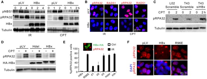
HBx impairs DNA end resection. (A) Immunoblotting for IR‐induced or CPT‐induced RPA32 and NBS1 phosphorylation in the presence or not of HBx. L02 cells were preinfected with 2 multiplicities of infection/cell of pLV (mock infection) or pLV‐HA‐HBx lentivirus. (B) pLV‐transfected or pLV‐HBx‐transfected L02 cells were treated as in (A), followed by immunostaining of RAD51 and pRPA32 foci. (C) Monitoring of RPA32 phosphorylation in indicated cells treated or not with siHBx. (D) Immunoblotting for pRPA32 in CPT‐treated L02 cells transfected with indicated expression plasmids. HBx and Hdel (HBx with H‐box deleted: amino acids 88‐98) were monitored by α‐HA. (E) Percentage of pRPA32 IRIF‐positive L02 cells pretransfected with different HA‐HBx constructs. pRPA32 foci were monitored 4 hours after irradiation. Cells with double‐positive staining of HA and phosphorylated serine 33 were judged as positive except in “HBx−” cells (no HA‐HBx transfection) where pRPA32 were counted in random cells. Inset: Costaining of HA‐HBx (green) and phosphorylated serine 33 (red). n = 3 biological repeats. Error bars = SD. (F) Immunofluorescence for pRPA32 in L02 cells infected with HBx wild‐type or R96E lentivirus (2 multiplicities of infection/cell) followed by IR treatment. Abbreviations: DAPI, 4′,6‐diamidino‐2‐phenylindole; HA, hemagglutinin; LV, lentivirus.