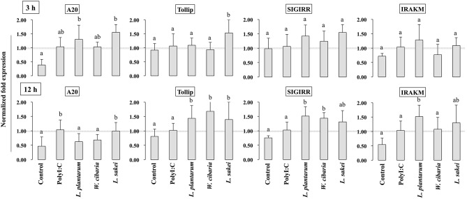
Figure 8.
Analysis of TLRs negative regulators expression in HCT116 cells. After HCT116 cells treated LAB strains and PolyI:C for 3 and 12 h. The expression of A20, Tollip, SIGIRR, and IRAK-M were evaluated by RT-PCR. Cells treated with either PolyI:C or medium alone were used as positive and negative controls, respectively. The positive control was used for comparison of LAB strains treated groups The mean differences among the different superscript letters (a, b, ab) were significant at 0.05 level. LABs were able to modulate expression of negative regulators of TLR signaling in HCT116 cells, stimulated with PolyI:C.

