
Figure 2:
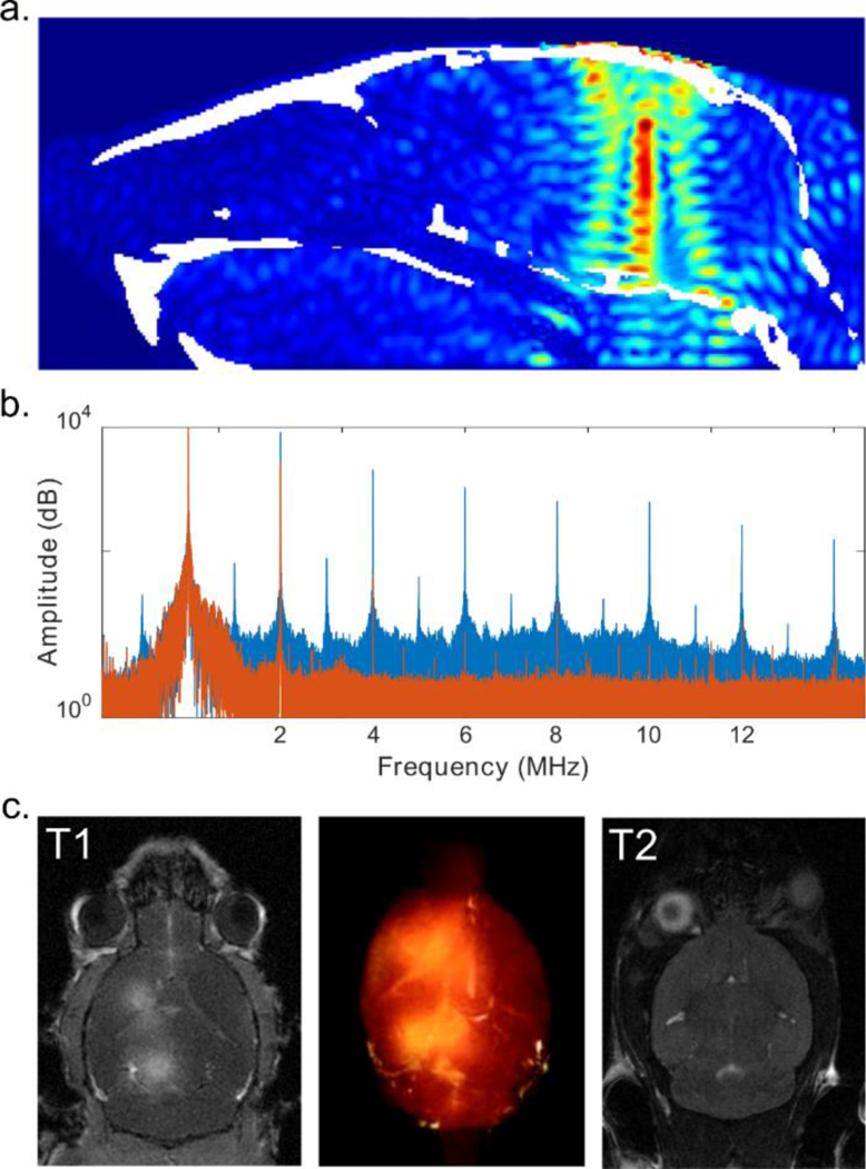
FUS-facilitated delivery. a. CT-based simulation revealed the FUS beam focus in situ (mouse skull is indicated in white). b. Passive cavitation detection was used to monitor the sonication in real-time without (orange) and with (blue) microbubbles. c. BBB opening was confirmed with contrast-enhanced T1-weighted MRI (left) and rendered in 3D (middle). No edema was observed on T2 MRI one-day post sonication.