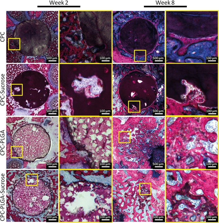
Figure 3.

Histological overview and magnifications of CPC formulations implanted in a rat femoral bone defect at two (left panels) and 8 weeks (right panels). Sucrose porogen and PLGA porogen or pores resulting from sucrose porogen dissolution (large) or PLGA porogen degradation (small) can be discriminated based on size differences. Large pores resulting from sucrose porogen dissolution are not homogeneously distributed. At 2 weeks, limited tissue infiltration in peripheral pores resulting from sucrose porogen dissolution is apparent. At 8 weeks, significant degradation of PLGA‐containing CPC formulations can be observed. Pure CPC shows hardly any degradation over the entire implantation period.
