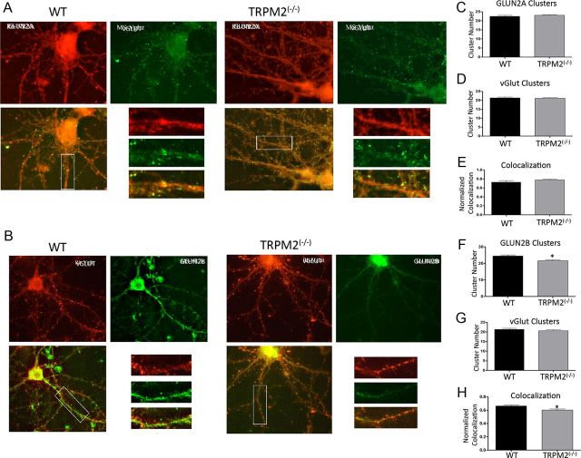
Figure 5.
TRPM2−/− reduces synaptic colocalization of GluN2B, but not GluN2A. Immunofluorescence of detecting GluN2A and GluN2B clusters in hippocampal CA1 neurons from WT and TRPM2−/− animals. A, To detect synaptic GluN2A, equal dendritic segments were used to count labeled clusters, where green fluorescence represents synaptic marker VGLUT expression and red represents GluN2A subunit expression. B, Synaptic GluN2B are labeled as follows: red fluorescence is VGLUT, and green is GluN2B. Overlaid images were used to count colocalized clusters. C, WT and TRPM2−/− had no difference in number of GluN2A clusters (n = 27; p > 0.05). D, E, There was also no change in number of synapse and GluN2A synaptic colocalization between neurons from WT and TRPM2−/− animals (n = 27; p > 0.05). F, The number of GluN2B clusters were reduced in hippocampal neurons from TRPM2−/− animals compared with those from WT animals (n = 24). *p < 0.05. G, H, GluN2B synaptic colocalization was also reduced in TRPM2-null neurons compared with WT (n = 24; *p < 0.05), with no effect on total number of synapses. H, Colocalization of GluN2B and VGLUT was reduced in TRPM2−/− compared with WT.

