Figure 2.
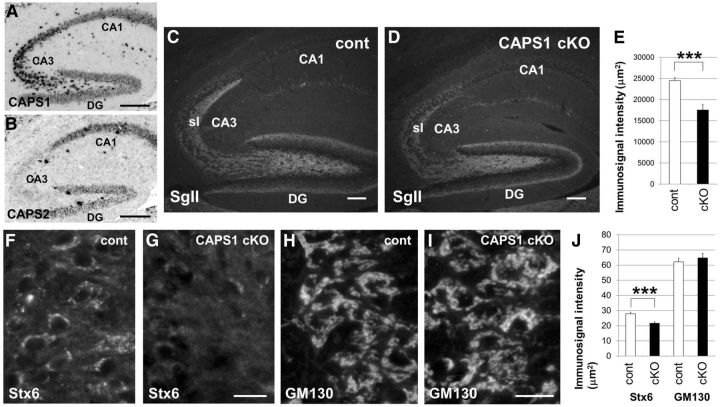
Stx6 immunolabeling was diffusely distributed in the soma in Caps1 cKO mice. A, B, Cellular localization of Caps1 (A) and Caps2 (B) mRNA by in situ hybridization in the hippocampus of P21 wild-type mice. Scale bar, 50 μm. C, D, Immunohistochemical localization of SgII in sagittal sections of the hippocampus in control (flox/−) (cont; C) and Caps1 cKO [flox/−, Emx1(+/−)] (D) mice at P21. sl, Stratum lucidum. Scale bar, 25 μm. E, SgII immunolabeling intensities in the stratum lucidum of control (white bar; 8 sections from 4 mice) and Caps1 cKO (black bar; 6 sections from 3 mice) mice are shown. Error bars indicate the SEM. ***p = 0.000302, by Student's t test. F–I, Sagittal sections of control (flox/−) (F, H) and Caps1 cKO [flox/−, Emx1(+/−)] (G, I) P21 hippocampus, showing CA3 pyramidal cells immunostained for Stx6 (F, G) and GM130 (H, I). Scale bar, 20 μm. J, Immunolabeling intensities in control (white bar) and Caps1 cKO (black bar) mice are shown. Stx6 (n = 70 for control and n = 70 for Caps1 cKO) and GM130 (n = 46 for control and n = 54 for Caps1 cKO) immunostaining was performed on seven sections from three mice. Error bars indicate the SEM. ***p = 0.0000289, by Student's t test.

