Figure 3.

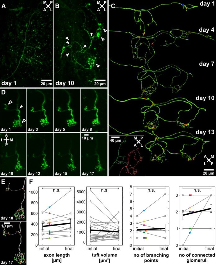
Repeated in vivo time-lapse imaging of single identified ORNs of larvae reveals no reduction of bifurcations or number of connected glomeruli during neuronal maturation. A, A single-labeled, immature ORN growing into the OB shows a number of branching points but lacks tufted arborizations characteristic for glomerular innervations. B, The same ORN after 10 d shows extensive axon growth with multiple branching points (filled arrowheads) and multiple established connections to at least two different glomeruli (open arrowheads). C, Three-dimensional reconstructions of multiple recordings of the same ORN depicted in A and B illustrate the process of axonal outgrowth and glomerular innervation (orange represents branching points). Inset, Comparison of the recordings on day 1 (green) and day 10 (red). D, Time series of a mature ORN with one branching point (filled arrowhead) and established connections to two different glomeruli (open arrowhead). The axonal growth pattern does not change over the observed time of 16 d. E, Three-dimensional reconstructions of the cell depicted in D (one week distance). Inside the glomeruli, the axon arborizes multiple times (orange represents branching points), and synaptic contacts characterized by axonal thickenings can be identified (green). Whereas the rough axonal growth pattern is retained, intraglomerular connections are refined. F, Quantitative comparison of axonal length, tuft volume, branching points, and connected glomeruli in the initial and the final state (n = 10). Error bars indicate SEM; statistical significance was tested using a paired t test. Same cells are indicated by colored dots. Mean initial and final values are connected by a black line. n.s., Not significant.
