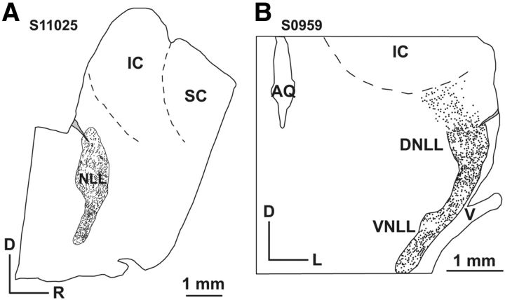
Figure 2.
Histological reconstruction of recording sites of two animals. Overlays of tracings of fluorescent images and cresyl violet stained sections. A, Caudal approach, parasagittal view. B, Lateral approach, coronal view. AQ, aqueductus; D dorsal; L, lateral; NLL, nuclei of the lateral lemniscus; R, rostral; SC, superior colliculus; V, trigeminal nerve. Electrode tracks are marked in light gray with black outlines. The VNLL and DNLL were first outlined; within those outlines locations of nucleated cell bodies are indicated with a dot (see main text).

