
Figure 7.
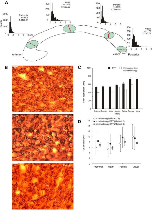
A, Distribution of axon diameters sampled from discrete dorsoventrally oriented probes in different anteroposterior sectors of the CC, where fibers from prefrontal, motor, posterior parietal, and visual cortex cross the midline. Data refer to subject 489-07. For each histogram, number of counts, relative mean diameter, and SD are provided. B, Microphotographs of myelin-stained callosal axons from prefrontal, motor, and parietal cortex. White circles are examples of axons sampled. Calibration, 10 μm. C, Comparison of the length of callosal axons from their origin to the CC midline as derived from DTT and by rescaling the length estimates from monkeys to the human brain volume (see Caminiti et al., 2009) in humans. D, Conduction delays to the CC midline computed by using information about axon diameters and conduction distance derived from DTT (methods 2 and 3) or by the rescaling procedure (method 1) described in the text.