Figure 1.
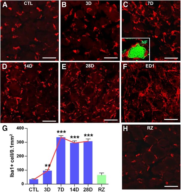
SNI increased the number of macrophages in the DRGs. A–E, Immunohistochemical detection of Iba1 expression in DRG sections. SNI was inflicted, and ipsilateral L4 and L5 DRGs were obtained at 3 (B), 7 (C), 14 (D), and 28 (E) d after SNI. The number of Iba1-positive macrophages was compared with that in control (CTL) DRGs from a sham operation (A). C, The inset shows a DRG neuron (green, stained with anti-neurofilament antibody) surrounded by Iba1-positive macrophages (red). F, Another macrophage marker, ED1, showed a similar level of expression at 7 d after SNI. G, Quantification graph of the number of Iba1-positive macrophages per 0.1 mm2 of DRG tissue. A red line was drawn to illustrate the temporal pattern of the SNI-induced macrophage increase. A green bar in the graph (G) represents the mean number of macrophages in the DRGs at 7 d after an injury to the central branch [rhizotomy (RZ)]. **p < 0.01 and ***p < 0.001 by one-way ANOVA, followed by Tukey's post hoc analysis. n = 3–5 animals for each group. H, DRG sections obtained at 7 d after rhizotomy were immunostained with Iba1 antibody. In contrast to the peripheral SNI, rhizotomy did not increase macrophage number. Scale bars, 50 μm.

