Figure 5.
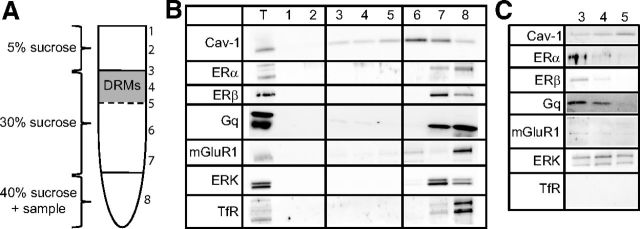
ERs and E2-sensitive signaling molecules are present in DRMs of the hippocampus. A, Depiction of the sucrose gradient used to fractionate hippocampal homogenates. DRMs formed a distinct band at the 5–30% sucrose interface. Samples were collected from the top down, and were subjected to Western blotting for Cav-1, ERα, ERβ, ERK, mGluR1, and TfR (negative control for DRMs). B, Western blots of 20 μg for each sample plus total lysate (T). With 1–2 min exposure, protein was observed in some fractions (T, 7, and 8), and light bands for Cav-1, ERK, and mGluR1 were also observed in DRM fractions. C, Hippocampal DRM fractions (20 μg) were run separately and probed as in B with 5–15 min exposure times. All proteins were visible, except for TfR.

