
Figure 1.
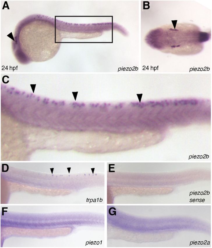
The expression pattern of piezo2b during zebrafish development. A–C, In situ hybridization analysis using an antisense piezo2b probe at 24 hpf. A, Piezo2b expression can be observed in the trigeminal ganglion (arrowhead) and Rohon–Beard cells (box). B, Dorsal view showing piezo2b expression in the trigeminal ganglion (arrowhead). C, Higher magnification of the boxed region in A. Piezo2b expression can be detected in the Rohon–Beard cells (arrowheads). E, To ensure that these observations were specific, we performed an identical assay using a sense piezo2b probe, which did not label anything. D, In situ hybridization analysis using an antisense trpa1b probe indicates that this gene is also expressed in Rohon–Beard neurons (arrowheads). F, G, The other two zebrafish piezo homologs, piezo1 and piezo2a, do not appear to be expressed in Rohon–Beard cells.