
Figure 1.
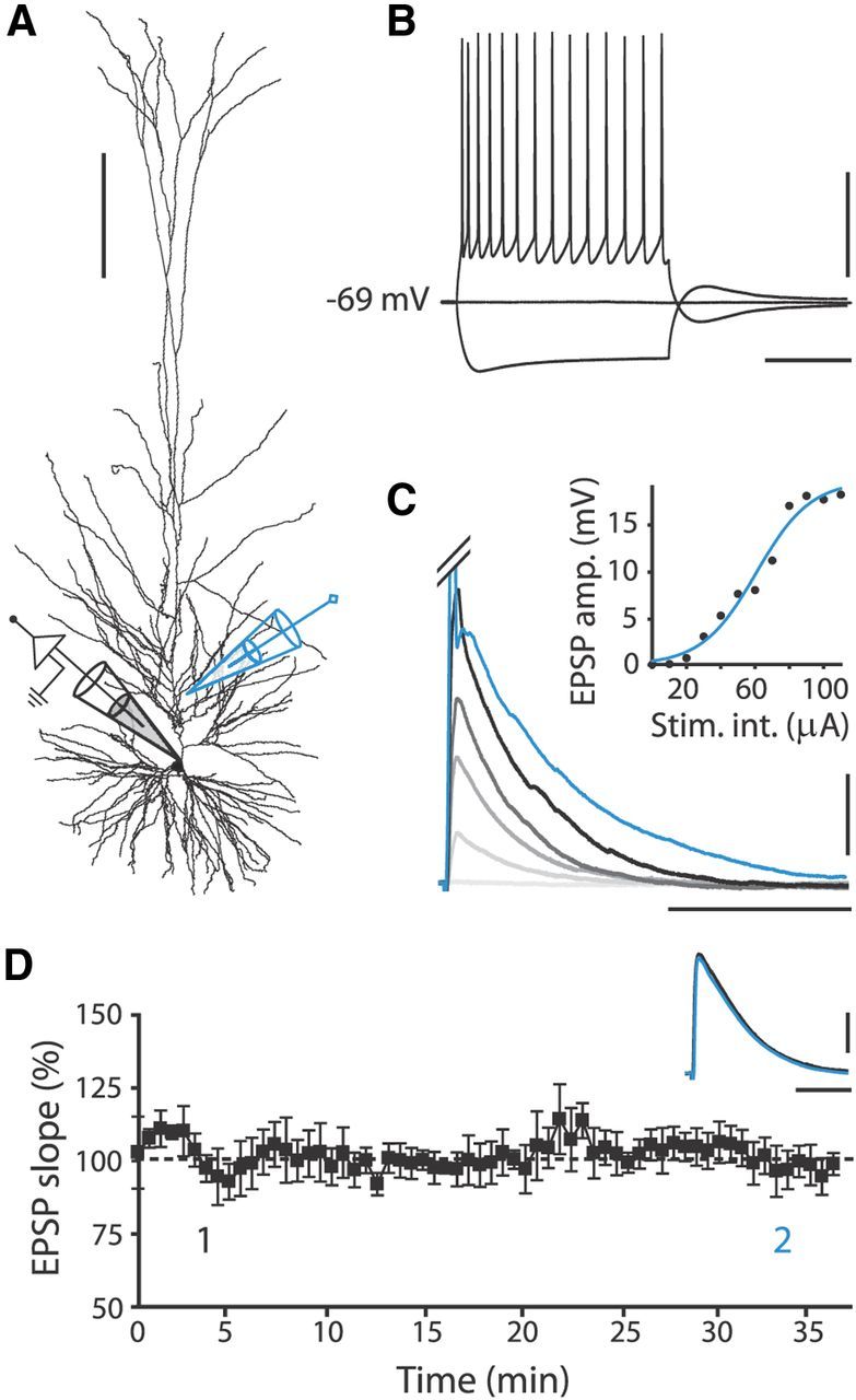
Recording pyramidal neurons in slices of human medial temporal cortex. A, Reconstruction of biocytin-labeled human pyramidal neuron, showing relative positions of recording (black) and stimulating (blue) electrode. Calibration: 200 μm. B, Typical voltage response to hyperpolarizing (−200 pA) and depolarizing (+250 pA) current steps (500 ms) of human pyramidal neuron. Calibration: 40 mV, 200 ms. C, Human EPSPs evoked by extracellular stimulation at different intensities starting at 10 μA, with 20 μA increments. Blue trace represents clipped AP evoked at 120 μA stimulation intensity (Stim. int.). Inset, Corresponding input/output curve. Calibration: 5 mV, 60 ms. D, EPSP slope over time normalized to mean EPSP slope in first 10 min of recording; no pairing to postsynaptic APs was applied. Black squares, 7 EPSP mean ± SEM. Inset traces, Example EPSPs recorded within first 5 min (1, black) and after 30 min (2, blue). Calibration: 2 mV, 40 ms.