
Figure 1.
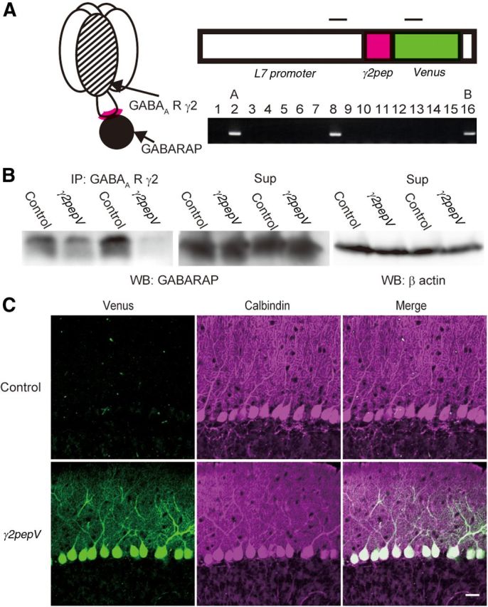
Expression of γ2pepV transgene in heterozygous mice. A, Schematic representation of binding of GABAA receptor γ2 subunit and GABARAP (left). Design of γ2pepV transgene (top) and its detection with PCR (bottom). Insertion of the transgene was examined in 16 mice, and it was detected in mice 2, 8, and 16. Top bars represent PCR primer target regions. The size of the PCR product was 468 base pairs. B, Interaction of GABARAP with GABAA receptor γ2 subunit in cerebellar extracts. The cerebellar extracts were coimmunoprecipitated with an antibody against the GABAA receptor γ2 subunit, followed by SDS-PAGE and Western blotting with an anti-GABARAP antibody. Wild-type (Control) mice showed stronger GABARAP signal than γ2pepV line A mice in the immunoprecipitate. GABARAP signal (middle) and β actin signal (right) in the supernatant are also shown. C, Cerebellar slices of control and line γ2pepV mice. Immunofluorescence for Venus (green) and that for calbindin (PN marker, magenta) are shown. Scale bar, 25 μm.