
Figure 2.
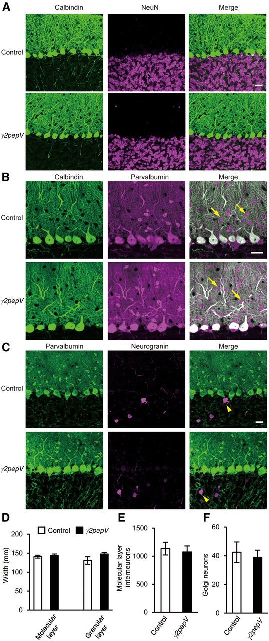
Morphology of the cerebellar cortex. Cerebellar slices stained with antibodies against calbindin (PN marker, green) and NeuN (granule neuron marker, magenta) (A), calbindin (green) and parvalbumin (a marker for molecular layer interneurons and PNs, magenta) (B), or parvalbumin (green) and neurogranin (Golgi neuron marker, magenta) (C). Arrows and arrowheads indicate molecular layer interneurons and Golgi neurons, respectively. Scale bars, 25 μm. D–F, The width of each layer (D) and the cell densities (cell number/mm2) of molecular layer interneurons (E) or Golgi neurons (F) are presented.