
Figure 1.
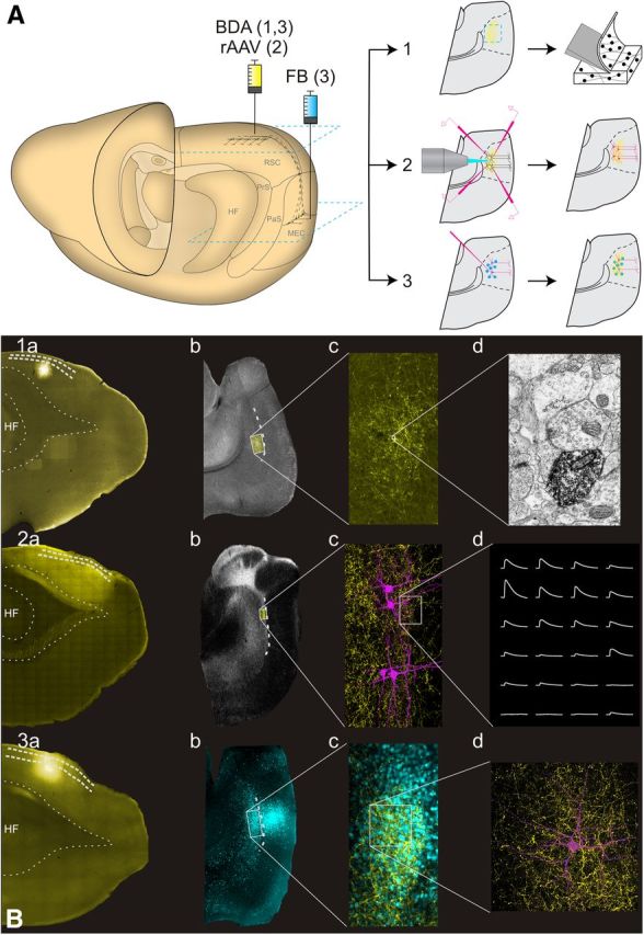
Experimental approaches used in the experiments. A, Schematic drawing of the rat brain and the injection sites (left). 1, The anterograde tracer BDA was injected into RSC for EM experiments. 2, rAAV was injected in RSC for optogenetic experiments. 3, The retrograde tracer FB was injected into superficial layers of MEC and BDA was injected into RSC for confocal experiments. The blue squares depict the level of the horizontal sections of the MEC and RSC shown in B. Each of the three experimental setups resulted in an anterogradely labeled plexus in MEC (yellow), intracellularly filled cells in Experiments 2 and 3 (magenta), and in Experiment 3 also retrogradely labeled neurons in MEC (blue dots). B, Examples of individual experimental protocols. 1, For EM experiments, BDA was labeled with AF546-conjugated streptavidin so that injections (1a) and an anterograde plexus (1b, 1c) could be identified (yellow). Sections were processed for EM, and synaptic complexes labeled with DAB were sampled in EM (1d). 2, For in vitro optogenetic experiments, mCherry fluorescence (yellow) was used for identifying the injection in RSC (2a), anterograde plexus in MEC (2b, 2c), where intracellular recordings and axonal reconstructions were performed (2c, 2d). 3, For confocal analysis, BDA was labeled with AF488 (yellow; 3a, 3c), and FB-positive cells (blue; 3b, 3c) were filled with AF568 (magenta; 3d). PaS, Parasubiculum; PrS, presubiculum.