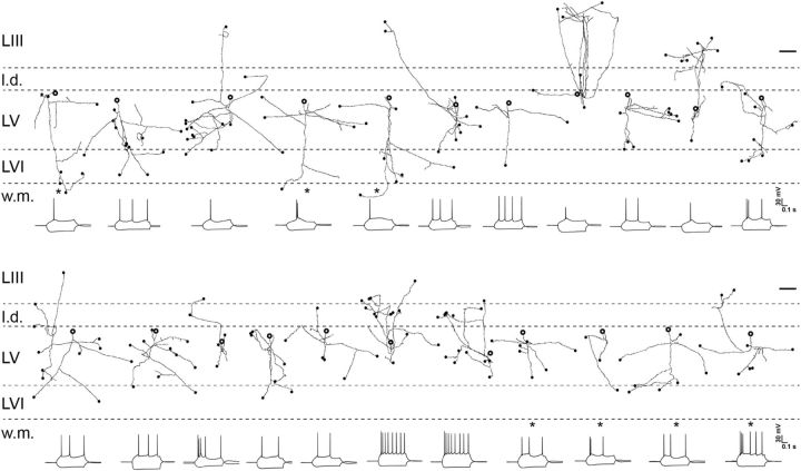
Figure 6.
Individual axonal reconstructions of 22 pyramidal cells recorded in horizontal slices through MEC. The one interneuron (Fig. 5D) is not included. The position of the soma in layer V is indicated with the bold open circle (size of the soma is not to scale). Axons that appeared to be cut at the surface of the slice are indicated by filled circles (scale bar, 200 μm). Below each neuron is the characteristic electrophysiological test response to current steps of ±100 pA (calibration: 0.1 s, 30 mV). The cells indicated with an asterisk are those for which the dendritic morphology is shown in Figure 5, B (4 cells in the bottom row) and E (3 cells in the top row). l.d., Lamina dissecans; L, layer; w.m., white matter.

