Figure 5.
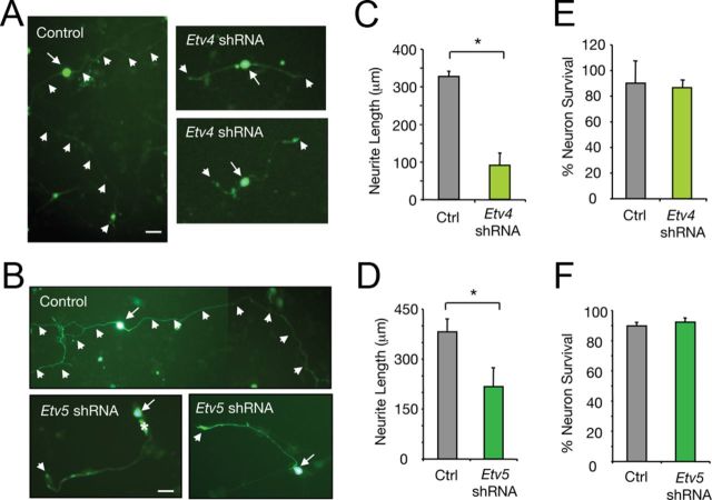
Etv4 and Etv5 are involved in NGF-induced sensory neuron differentiation. A, B, Dissociated DRG neurons transfected with control, Etv4-shRNA (A), or Etv5-shRNA (B) expressing GFP constructs and maintained in the presence of NGF (50 ng/ml). After 36 h in culture, neurons were fixed and stained with anti-βIII-tubulin antibodies (data not shown). Scale bars, 20 μm. Arrows indicate neuronal CBs and arrowheads indicate neurite trajectory and neurite tips. C, D, Histograms show the inhibition of neurite outgrowth in DRG neurons by knockdown of Etv4 (C) or Etv5 (D) expression. The length of the longest neurite was measured. Results are shown as the average ± SD of a representative experiment performed in triplicate. C, *p = 0.0135 (Student's t test, t = 4.2); D, *p = 0.0003 (Student's t test, t = 11.6). The experiment was repeated at least two times with similar results. E, F, Histograms showing the survival of DRG neurons transfected with control, Etv4-shRNA (E) or Etv5-shRNA (F). Neuronal survival was evaluated using DAPI for the nuclear staining. GFP-positive neurons containing fragmented or condensed nuclear staining were scored as apoptotic cells. The results are averages ± SD of a representative experiment performed in triplicate.

