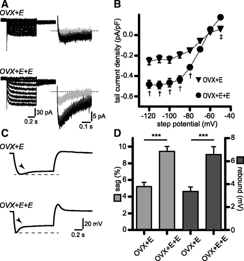
Figure 5.

Upregulation of Ih and associated sag and rebound is mimicked by high circulating estradiol levels in a luteinizing hormone-surge protocol. A, Membrane currents in OVX+E and OVX+E+E RP3V kisspeptin neurons in response to voltage steps from −50 to −120 mV in the presence of a cocktail of synaptic and ion channel blockers (see Results). Steady-state (left) and tail currents (right) were larger in OVX+E+E. Only tail currents corresponding to −70 (light gray), −90 (dark gray), and −110 mV steps (black) are illustrated for clarity. B, Corresponding tail current activation curves illustrating the upregulation of Ih in OVX+E+E. OVX+E: n = 18 neurons, 3 mice; OVX+E+E: n = 17 neurons, 3 mice. †p < 0.001, ‡p < 0.05 two-way ANOVA with Bonferroni post-test. C, Example traces illustrating the depolarizing sag and rebound seen in OVX+E and OVX+E+E RP3V kisspeptin neurons in response to the hyperpolarizing current injections. Arrowheads indicate the depolarizing sag. D, Histogram summarizing the sag and rebound amplitudes in OVX+E and OVX+E+E. OVX+E: n = 18 neurons, 3 mice; OVX+E+E: n = 17 neurons, 3 mice; ***p < 0.001.
