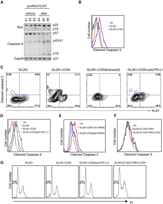
Figure 5.

Neuronal PD-L1 kills glioma cells via caspase-dependent apoptosis. A, GL261 cells (CFSE labeled) purified with FACS after coculture with CGNs. Western blot of BAX, caspase 8, and GAPDH in GL261 cells before and after coculture with CGNs. GAPDH was the loading control. B, FACS histogram of cleaved caspase 3 in GL261 cells before and after coculture with CGNs (gated on CFSE). C, FACS staining for Ki-67 and caspase-3 in GL261 cells without and after coculture with 3 DIV CGNs (or blocking with anti-PD-L1 antibody). GL261 cells and CGNs were separated with a transwell membrane. One representative of three experiments is shown. D, Cleaved caspase 3 in gated GL261 cells before and after coculture with CGNs; anti-PD-L1 (20 μg/ml) was applied to CGNs for 30 min before coculturing. E, Cleaved caspase 3 in gated GL261 cells before and after coculture with control-transfected CGNs (ctrl siRNA) or pdl1-silenced CGNs (pdl1 siRNA). F, Caspase inhibitor (Z-VAD-FMK, 20 μm) was added to GL261 cells 2 h before coculturing. G, PI staining of GL261 cells before and after coculture with CGNs, with anti-PD-L1 added to CGNS or Z-VAD-FMK to GL261 cells.
