
Figure 9.
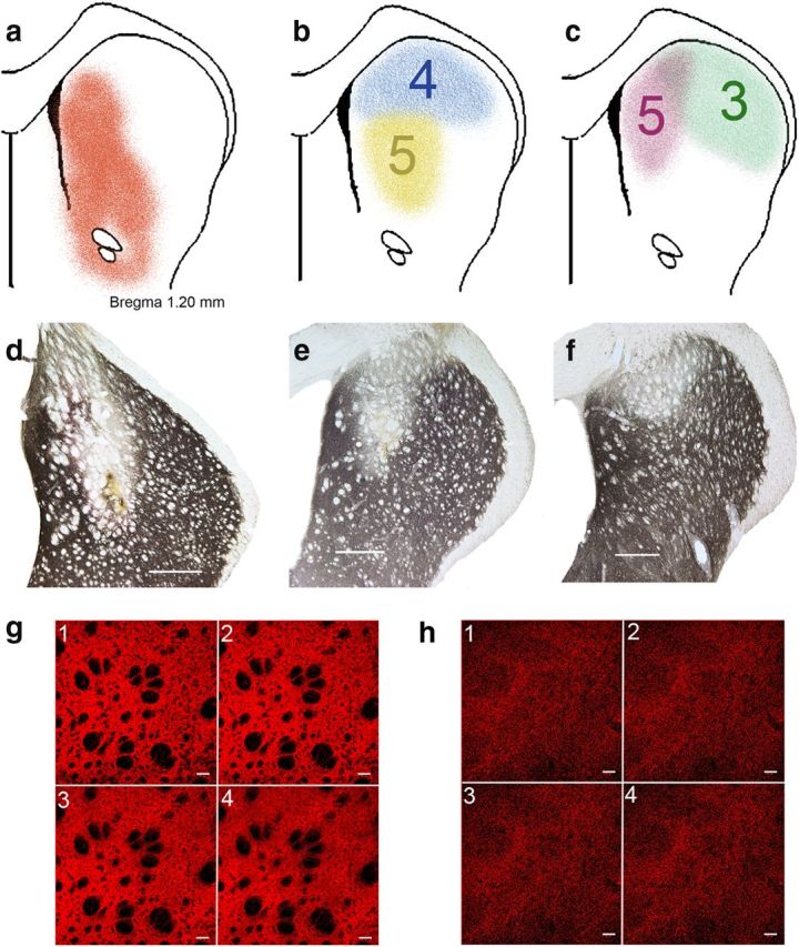
6-OHDA-induced loss of TH immunoreactivity in the caudate nucleus. a, Striatal projection region of the medial prefrontal cortex, which was the target region for infusions of 6-OHDA (reproduced from Mailly et al., 2013, their Fig. 4c). b, c, Representative extensions of loss of TH immunoreactivity and rating of placements (5 [best] to 1; see Materials and Methods). The yellow and pink lesions received highest ratings for placement, whereas the deafferentation of the blue and green examples was only partly placed in the target region and thus received lower ratings. d–f, Sections showing TH immunoreactivity in the caudate of lesioned hemispheres. Scale bars, 500 μm. To further exemplify the ratings of these lesions, d was rated 5 for placement and 5 for size (for definition, see Materials and Methods), e was rated 5 for placement and 2 for size, and f received a 4 for placement and a 2 for size. g, h, TH immunofluorescence in the caudate nucleus (confocal microscopy) of an intact rat (g; 1–4 are consecutive slices; 4.71 μm/slice) and a 6-OHDA-lesioned rat (h; 3.77 μm/slice; scales in slices are 40 μm). Morphometric analyses of residual TH puncta indicated a consistently near complete (>90%) reduction of TH immunofluorescence in the center of 6-OHDA lesions. In DL rats, the placement and size of the dopaminergic lesions correlated with fall rates.