
Figure 2.
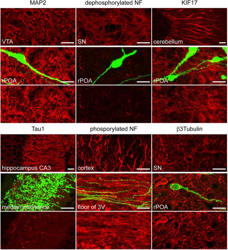
GnRH neuron processes do not label for classical axonal or dendritic markers. All markers (red label) labeled appropriate neuronal compartments in various other brain areas (top) but did not localize to GnRH neurons (green, middle). The bottom part of the same field, without GnRH GFP, is given for each marker to show that it is not expressed by GnRH neurons. Dendritic markers used were microtubule-associated protein 2 (MAP2) and dephosphorylated neurofilament and kinesin-like protein 17 (KIF17). Axonal markers were Tau1 and phosphorylated neurofilaments (NF). The general neuronal marker β-3-tubulin did not label GnRH neurons. 3V, third ventricle; rPOA, rostral preoptic area; SN, substantia nigra; VTA, ventral tegmental area. Scale bars: 10 μm.