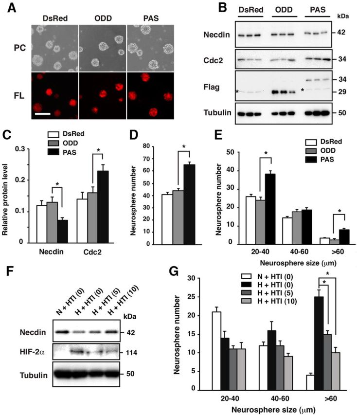
Figure 8.

The HIF-2α PAS domain downregulates endogenous necdin level and facilitates NSC proliferation. A, Lentivirus-infected NSCs. NSCs were infected with lentiviruses expressing DsRed (DsRed, empty vector), Flag-tagged HIF-2α PAS domain (PAS), and Flag-tagged ODD (ODD), and cultured for 6 d. Images of phase-contrast (PC) and fluorescence (FL) of lentivirus-infected NSCs are shown. Scale bar, 100 μm. B, C, Expression of necdin and Cdc2 proteins. Lysates of infected NSCs were analyzed by Western blotting using antibodies to necdin, Cdc2, Flag, and β-tubulin (B). Asterisks indicate nonspecific bands. The protein levels were quantified and normalized to β-tubulin (C). D, E, Neurosphere assay. Lentivirus-infected CD133+ NSCs were subjected to neurosphere assay in normoxia. The total number of neurospheres (>20 μm in diameter; D) and the numbers of three groups (20–40, 40–60, >60 μm in diameter) were counted (E). F, G, Effects of HIF-2α translation inhibitor. Primary NSCs were treated with HIF-2α translation inhibitor (HTI) at 0, 5, and 10 μm for another 3 d in hypoxia (H). Control NSCs were cultured for 9 d in normoxia (N). HIF-2α and necdin were analyzed by Western blotting. G, Neurosphere assay. Primary NSCs were subjected to neurosphere assay in the presence of the translation inhibitor. The numbers of three groups were counted. C–E, G, mean ± SEM (n = 3). *p < 0.05.
