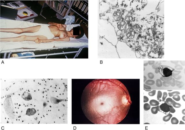
Figure 2.
Clinical and morphological observations in gangliosidoses. A, Near end stage in a Spanish 13-year-old patient with GM2-gangliosidosis variant B1 (photograph courtesy of H.H. Goebel, Mainz). B, GM2-gangliosidosis variant 0 in a 23-week-old fetus; and part of a cortical neuron; early stage of membranous cytoplasmic bodies (two clusters marked by crosses), i.e., lipid storing secondary lysosomes, ×36,000 (courtesy W. Schlote, Tübingen). C, The large cells are neurons distended by lysosomal storage in the medulla oblongata of a 1.5-year-old patient with GM1-gangliosidosis. The small dark bodies are nuclei from increased numbers of glial cells (“reactive gliosis”). Cresyl violet stain ×300 (courtesy of H.U. Benz, Tübingen). D, Cherry red macular spot in an infantile patient with GM2-gangliosidosis variant B (Tay–Sachs disease). The dark red spot in the middle of the light central area is secondary to lipid storage in neuronal cells in this area; the storing cells have lost their processes that normally cover the fovea centralis. The fovea normally appears as yellow, but is changed to the red macular spot showing the color of the choroidea behind the retina. E, Lymphocytes with clusters of vacuoles (storage lysosomes) in blood smear of an early infantile patient with GM1-gangliosidosis, indicating the “generalized storage disorder.” May-Grünwald stain; ×1500 (courtesy of H.U. Benz, Tübingen).

