
Figure 2.
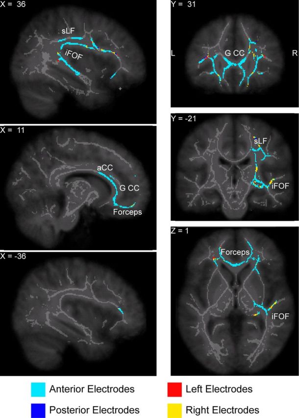
White matter tracts where axial diffusivity was higher in individuals with a steeper slow-wave slope. The plot shows the voxels with a positive significant correlation (thresholded at p ≤ 0.05 after correction for multiple comparisons) superimposed on the white matter skeleton (light gray) and on the study-specific MRI template (dark gray) following the neurological convention (right is right). The average slope was computed from the slow waves detected in all the four regions of interest, while the color coding indicates which region of interest was the most correlated with the AD at each voxel. The white matter tracts in which AD was associated with the slope steepness are concentrated in the frontal white matter, including the forceps minor, the genu and anterior part of the corpus callosum (G CC and aCC, respectively). These frontal pathways connect to the superior longitudinal fascicle (sLF) and the temporal branch of the inferior fronto-occipital fascicle (iFOF).