
Figure 3.
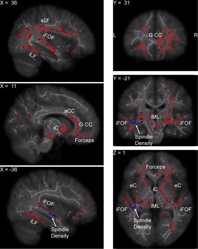
White matter tracts where axial diffusivity was higher in individuals with a stronger spindle power in sleep stage N2. Some of the white matter tracts in which AD was associated with spindle power partially overlap with the tracts plotted in Figure 2, such as the forceps minor, the genu and anterior part of the corpus callosum (G CC and aCC, respectively), the superior longitudinal fascicle (sLF), and the inferior fronto-occipital fascicle (iFOF). The network of white matter tracts whose AD was correlated with spindle power was more extended in the temporal lobe than the network where AD was associated with the slow-wave slope and included the inferior longitudinal fascicle (iLF) as well. Other tracts more selectively associated with spindle power include the white matter regions around the thalamus, such as the internal and external capsule (iC and eC, respectively) and the internal medullary lamina (iML) of the thalamus. In addition, tracts in the middle temporal lobe, colored in blue, were associated with spindle density in sleep stage N2.