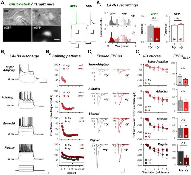
Figure 4.
Excitatory transmission onto amygdala interneurons is preserved in il1rapl1-deficient mice. A, Amygdalar GABAergic neurons were directly visualized and recorded by GFP fluorescence after crossing il1rapl1 mutant mice with GAD67-eGFP mice (see Materials and Methods). A1, Principal cells can be separated from interneurons by looking at cellular capacitance during the seal test. A2, Density and capacitance of GABA-ergic (GFP+) and principal (GFP−) cells in Il1rapl1 WT and KO preparations. Number of recorded cells is indicated. B, Spiking patterns of LA interneurons. B1, LA interneurons were classified in four subclasses based on spiking behavior (for a detailed description of interneuron classification, see Materials and Methods). B2, Mean spiking frequency against spike number for each subclass of interneuron. C, Excitatory evoked transmission of LA interneurons after thalamic stimulation. C1, Mean EPSC amplitude for 0.5, 1, and 5 mA stimulations in WT and KO interneurons. Calibration: 100 pA, 20 ms. C2, Left, I/O curves of LA interneurons for a 5 mA stimulation in WT and KO interneurons. Right, Mean EPSC amplitude at 5 mA stimulation intensity for all LA interneurons. Number of recorded cells is indicated.

