Figure 1.
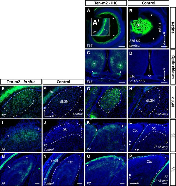
Ten-m2 is expressed in interconnected regions of the developing mouse visual system. Expression studies revealed the presence of Ten-m2 at multiple levels of the developing mouse visual system. Consistent expression patterns were seen using both in situ hybridization (E,F,I,J,M,N) and immunohistochemistry (A–D,G,H,K,L,O,P) in visual centers. A, B, Immunostaining for Ten-m2 in the retina was present in the RGC layer, including the region corresponding to the passage of fibers (arrowheads). By E16 expression was uniform across the dorsoventral retinal axis. At P0, immunostaining appeared more prominent at the junction of the IPL and RGC layer (arrow in A′). A high degree of nonspecific staining of the lens (asterisk) was seen (B; negative control from Ten-m2 KO mouse), although no staining was present in the retina of control sections. C, D, Near to the OC, Ten-m2 expression was observed on axonal tracts traveling to/away from the OC (arrowheads). No expression of Ten-m2 was seen on cells surrounding the chiasm where RGC axons travel (arrow). Areas that were dorsal to this region, corresponding to areas within the medial preoptic nucleus of the hypothalamus (asterisk), also showed immunoreactivity for Ten-m2. E–H, Uniform Ten-m2 expression in the dLGN (outlined with dashed line) was observed across the dorsomedial–ventrolateral axis (P7) with in situ hybridization (E). No staining was present in a nearby sense control section (F). Immunohistochemistry for Ten-m2 (G) shows a similar pattern to mRNA. No staining was present in sections incubated without primary antibody (H). I–L, The developing SC also displayed consistent expression across its retinorecipient layers (outlined by arrowheads) as shown by in situ hybridization (I) and immunostaining (K). No staining is present in the corresponding control sections (J, L). M–P, Deep layers of primary visual cortex (V1) expressed Ten-m2 mRNA by P0 (outlined by arrowheads). No staining is present in sense controls (N). By P7, immunohistochemistry reveals high expression in layers IV and V (area outlined by arrowheads) (O). No staining is seen in an adjacent control section (P). Images are representative coronal sections with dorsal to the top. Medial is to the right in all images except C and D, where medial is in the center. Dashed outlines in control images indicate areas of interest. Ctx, cortex; hc, hippocampus. Where applicable, the midline is indicated with a dotted line. Scale bars: 200 μm.

