
Figure 3.
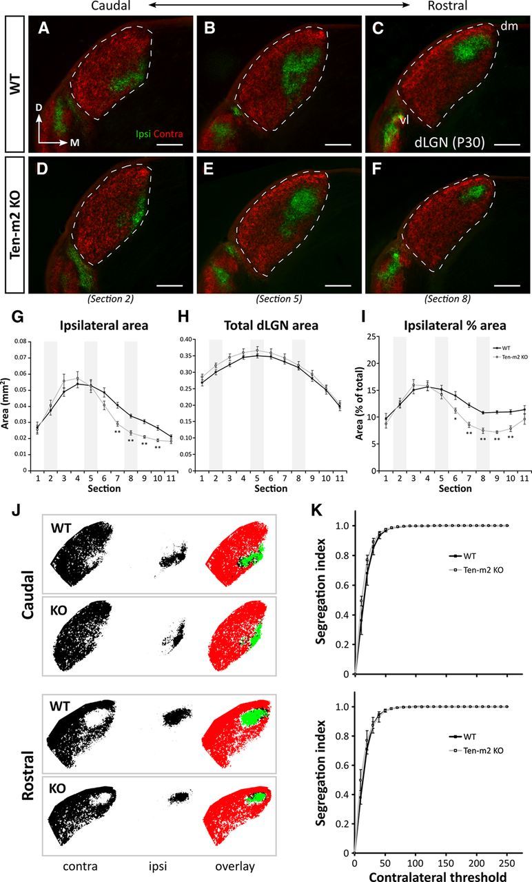
Retinogeniculate projections are distinctly altered in Ten-m2 KO mice. A series of caudal-to-rostral coronal sections from WT mice at P30 (A–C) demonstrate the stereotypical position of ipsilateral (green) retinal projections terminating within dLGN (outlined), segregated from contralateral (red) inputs, following dual intraocular injections of fluorescently labeled CTB. No difference in segregation or the overall placement of ipsilateral label was found between WT (n = 18 dLGNs) and Ten-m2 KO (n = 16 dLGNs; D–F) mice. An overall decrease in ipsilateral projections was found for Ten-m2 KO mice, which was seen predominantly within rostral dLGN (F vs C). In all images, dorsal is to the top and medial to the right. dm, Dorsomedial; vl, ventrolateral. Scale bar, 200 μm. G–I, Quantitative analysis of the caudal-to-rostral series (sections 1–11) through the dLGN revealed that absolute area occupied by ipsilateral terminals (G) was significantly reduced in a number of sections (7–10) through rostral Ten-m2 KO dLGN (light gray), compared with WT (black). These changes occurred in the absence of significant differences in total dLGN size (H), and were similarly observed when ipsilateral area was considered as a percentage of total dLGN area (I). Representative images shown in A–E correspond to sections 2, 5, and 8 along the caudal-to-rostral series in G–I (shaded areas). *p < 0.05, **p < 0.005, in comparisons to WT; multivariate ANOVA. J, Segregation of contralateral (left, red) and ipsilateral (middle, green) inputs to the dLGN was assessed in caudal (top) and rostral (bottom) dLGN using a threshold analysis. Overlapping contralateral and ipsilateral regions are indicated as black in image overlays (right). K, At each contralateral threshold value, segregation indices were not significantly different (p > 0.05) between WT (black) and Ten-m2 KOs (light gray) for either caudal (top) or rostral (bottom) locations, indicating comparable segregation of inputs in these animals.