Figure 5.

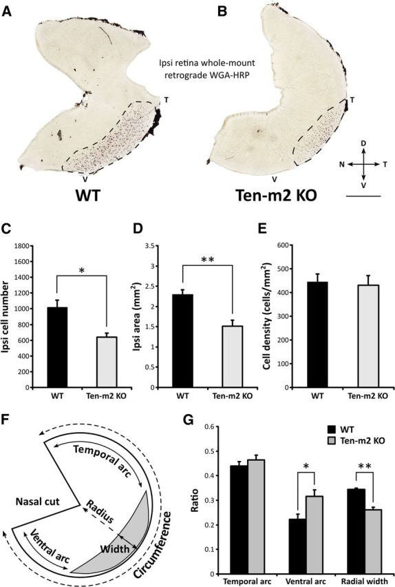
Retrograde tracing of ipsilateral retinogeniculate projections reveals distinct changes in Ten-m2 KO mice. Ipsilateral retinogeniculate projections were retrogradely labeled with stereotaxic WGA-HRP injections into the right dLGN. A, B, Labeled cells (dark spots; area outlined by dotted line) were visualized in whole mounts of ipsilateral retinae, with cells in WT (n = 5) found in a stereotypical position in VT retina (A). Both the number (C) and area occupied (D) by ipsilateral cells were significantly reduced in Ten-m2 KO retina (n = 4) (B). No differences were found in cell density (E) between genotypes. F, Schematic diagram outlining analysis of changes in the population of ipsilaterally labeled cells. Using the nasal fiducial cut, temporal and ventral arc length was expressed as a ratio of retinal circumference. The width of the ipsilateral label was expressed as a ratio of length across the radius of the retina. G, Ventral arc ratio was significantly increased in Ten-m2 KO retina, indicating the loss of ipsilaterally projecting cells from ventral retina, while no change in temporal arc ratio was found. Furthermore, a reduction in the ipsilateral radial ratio in Ten-m2 KOs indicated that changes also occur along the radial dimension of temporal retina. Dorsal is to the top, temporal to the right in all images. Scale bar, 1 mm. *p < 0.05, **p < 0.01, in comparisons to WT; Student's unpaired t test.
