Figure 6.

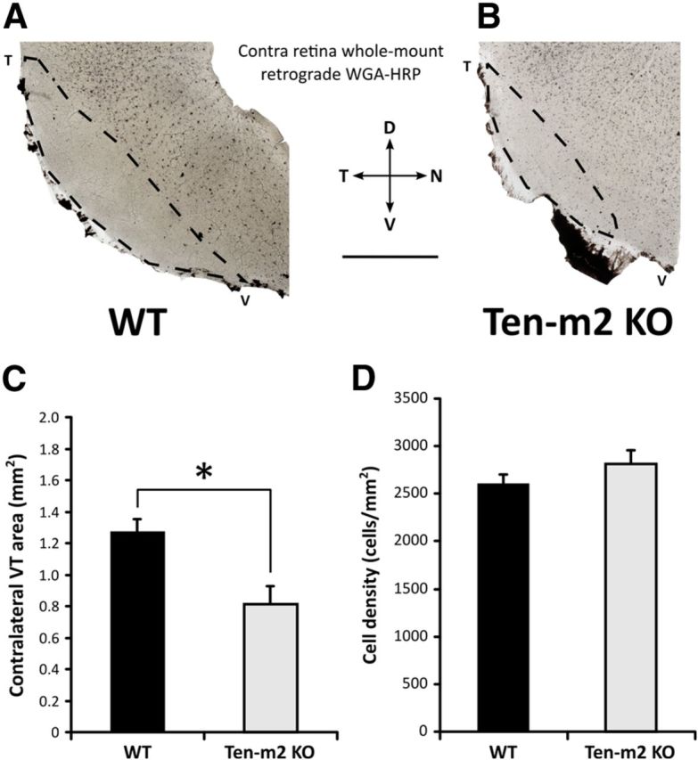
Ten-m2 KO mice display corresponding changes to contralateral retinogeniculate projections from VT retina. Retrograde tracing of contralateral retinogeniculate projections was achieved using stereotaxic WGA-HRP injections into dLGN. A, B, The VT quadrants of whole-mounted retinae, contralateral to the injections are shown. Labeled cells fill the bulk of this region, however, the VTC region, corresponding to the origin of ipsilateral projections (outlined), was largely devoid of labeled cells. The area of the sparsely labeled contralateral VT region (C) was significantly reduced in Ten-m2 KO mice, due to an expansion of retrogradely labeled contralaterally projecting cells into the ventral part of the VTC (B). The density of labeled cells within the central region of the retina was not significantly different across genotypes (D). Scale bar, 1 mm. Dorsal (D) is to the top, temporal (T) to the left, nasal (N) to the right, ventral (V) to the bottom. *p < 0.05, Student's unpaired t test.
