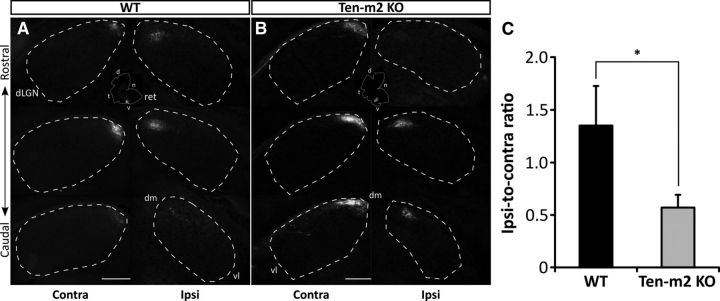
Figure 8.
Reduced ipsilateral and expanded contralateral projections from ventral retina map with normal topography in Ten-m2 KO mice. Focal injections of DiI were made into ventral retina (dotted outline; inset) of WT (A) and Ten-m2 KO (B) mice at P13. In both WT and Ten-m2 KOs, contralateral TZs were located within the dorsomedial pole of dLGN (dashed line), while ipsilateral TZs were displaced ventrolaterally. In Ten-m2 KOs, ipsilateral label was consistently reduced, while label appeared expanded in the contralateral dLGN. Despite this expansion, only a single patch of contralateral terminals in the topographically appropriate position at the dorsal margin of the dLGN was observed. C, Upon quantification, the ratio of ipsilateral-to-contralateral label showed a significant decrease in Ten-m2 KOs (n = 3), compared with WTs (n = 5). ret, retina; dm, dorsomedial; vl, ventrolateral; d, dorsal; t, temporal; n, nasal; v, ventral. Scale bar, 200 μm. *p < 0.05, Mann–Whitney U test.

