
Figure 1.
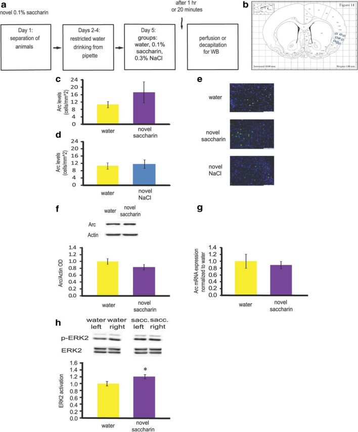
Bi-hemispheric Arc/Arg3.1 is not modulated in the gustatory cortex after novel taste learning. a, Schematic representation of the behavioral paradigm of novel taste learning. b, Rat coronal brain section containing the gustatory cortex with illustration of the nine sampling rectangles. c, d, Arc/Arg3.1 protein levels remain stable 1 h following novel taste learning for combined left and right hemispheres, as measured by immunohistochemistry. Rats drank 0.1% saccharin (n = 8) or 0.3% NaCl (n = 3) as a novel taste, or water (n = 7) as a familiar taste. p > 0.05. e, Immunofluorescence representative images (blue, Hoechst nuclear staining; green, Arc/Arg3.1 protein). Scale bar, 100 μm. f, Arc/Arg3.1 protein levels remain stable 1 h following novel taste learning for combined left and right hemispheres, as measured by Western blot. Rats drank 0.1% saccharin (n = 7) or water (n = 7) as a familiar taste. Top, Representative immune-blots of Arc/Arg3.1and Actin. p > 0.05. g, Arc mRNA levels remain stable 1 h following novel taste learning for combined left and right hemispheres, as measured by qRT-PCR. p > 0.05. h, When combining the volume of each band (area of the band * optical density) for left and right, ERK was activated 20 min following novel taste learning, as measured by Western blot. Rats drank 0.1% saccharin (n = 5) or water (n = 5) as a familiar taste. Top, Representative immune-blots of p-ERK and ERK proteins. *p < 0.05. Results represent mean ± SEM.