
Figure 13.
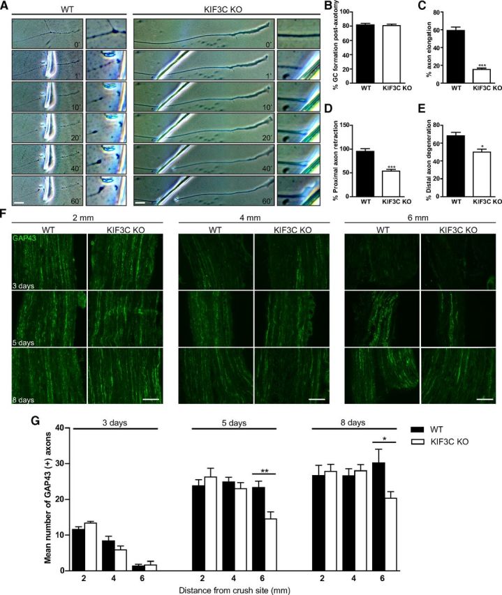
KIF3C is necessary for adult axon regeneration after injury. A, Successive frames from a time-lapse recording illustrating the behavior of a WT or KIF3C KO axon after axotomy in vitro. The enlarged box represents the site of axotomy at higher magnification. Scale bar, 20 μm. B, The percentage of WT or KIF3C KO axons that regrew growth cones after axotomy (n = 30 and n = 30, respectively, from three independent experiments). C, The percentage of WT or KIF3C KO axons that elongated after axotomy (n = 30 and n = 30, respectively). D, The percentage of WT or KIF3C KO axons that retracted after axotomy (n = 30 and n = 30, respectively). E, The percentage of WT or KIF3C KO distal axons that degenerated after axotomy (n = 30 and n = 30, respectively). Error bars indicate SD. *p < 0.05. ***p < 0.001. F, Representative photomicrographs of GAP43-positive axons in longitudinal cryosections from WT and KIF3C KO sciatic nerves, 3, 5, and 8 d after crush injury. Distance from crush site is 2, 4, and 6 mm. Scale bar, 100 μm. G, GAP43 axon count at 3, 5, and 8 d after sciatic nerve crush injury. Axon counts were taken at 2, 4, and 6 mm from the crush site. Error bars indicate SEM. *p < 0.05. **p < 0.01.