
Figure 3.
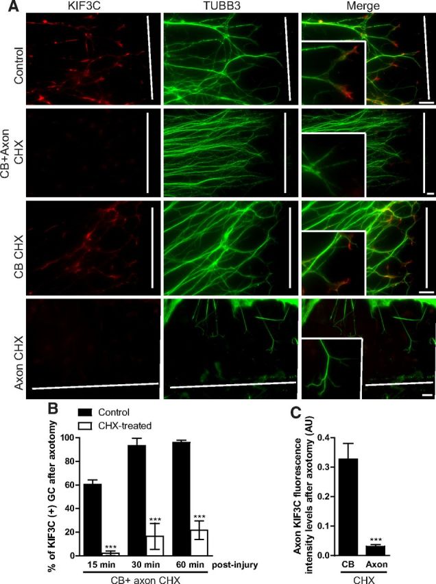
KIF3C is locally translated in embryonic axons after axotomy. A, Immunostaining of axotomized embryonic DRG axons cultured in Campenot chambers to separate the cell body (CB) from the axonal (Axon) compartment. The cultures were treated for 1 h with control medium or with the protein synthesis inhibitor cycloheximide (25 μm) (CHX), which was added to the CB and the axonal compartment, the CB compartment alone, or the axonal compartment alone. The white lines indicate the site of injury. B, Quantification of the percentage of growth cones (GC) enriched with KIF3C at different time points after axotomy. CHX was added to both the somatic and axonal compartments (30 axons were analyzed per condition and time point). C, Quantification of KIF3C fluorescence intensity levels in the growth cone of axotomized embryonic DRG axons. CHX was added either to the somatic or the axonal compartment (n = 30 and n = 30, compiled from three independent experiments). Error bars indicate SD. ***p < 0.001. Scale bar, 20 μm.