Figure 8.
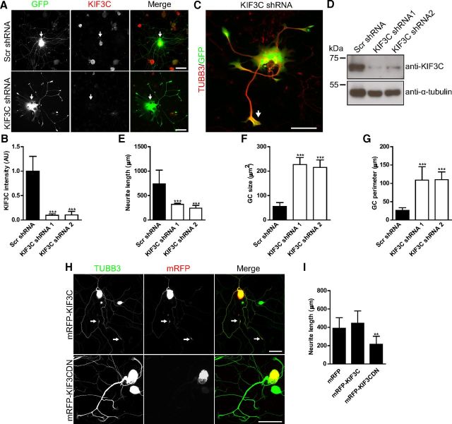
KIF3C knockdown in adult neurons impairs neuronal morphology and neurite outgrowth. A, Immunofluorescence images of control (Scr shRNA) and KIF3C-depleted (KIF3C shRNA) adult DRG neurons stained for KIF3C. Arrows indicate KIF3C staining in the cell bodies. Scale bar, 100 μm. B, Quantification of KIF3C fluorescence intensity levels in the cell bodies of adult DRG neurons transfected with control or KIF3C knockdown (KIF3C shRNA 1 or KIF3C shRNA 2) constructs (n = 98, n = 97, and n = 75, respectively, compiled from three independent experiments). C, Immunofluorescence image of an adult DRG neuron depleted of KIF3C and immunostained for TUBB3. The arrows indicate the enlarged growth cones. Scale bar, 100 μm. D, Cortical neurons were transfected with the indicated shRNAs and were lysed 24 h after transfection. Whole-cell lysates were probed for KIF3C or α-tubulin. E, Quantification of neurite length in adult DRG neurons transfected with control or KIF3C knockdown constructs (n = 114, n = 124, and n = 102, respectively, compiled from three independent experiments). F, Quantification of growth cone size in control and KIF3C knockdown neurons (n = 120 and n = 144, respectively, compiled from three independent experiments). G, Quantification of growth cone perimeter in KIF3C knockdown neurons (n = 120 and n = 144, respectively, compiled from three independent experiments). H, Immunofluorescence images of adult DRG neurons transfected with mRFP-KIF3C or mRFP-KIF3C dominant-negative (DN) cDNA and immunostained for TUBB3. mRFP-KIF3C accumulates at the growth cones as observed with the endogenous protein (arrows). Scale bar, 100 μm. I, Quantification of neurite length in control (mRFP), mRFP-KIF3C, or mRFP-KIF3CDN-transfected neurons (n = 158, n = 166, and n = 165, respectively, compiled from three independent experiments). Error bars indicate SD. **p < 0.01. ***p < 0.001.

