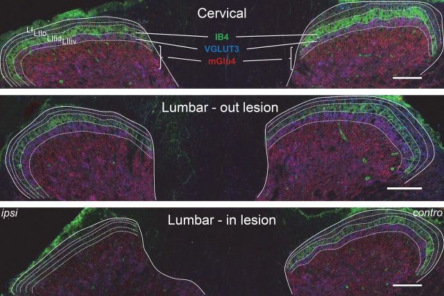
Figure 3.
Partial reduction of mGlu4 receptor staining in the dorsal horns of the spinal cord after rhizotomy. A cervical and two lumbar transverse sections of the spinal cord of a C57BL/6 mouse subjected to unilateral dorsal rhizotomy of the fourth lumbar nerve are displayed. Triple staining was performed against mGlu4 receptors (red), VGLUT3 (blue), and IB4 (green). The solid lines indicate the separation between gray and white matter; the dotted lines indicate lamina I (LI), outer lamina II (LIIo), and the dorsal and ventral inner lamina II (LIIid and LIIiv). Fifteen days after the operation, immunoreactivity of mGlu4 receptors is partially reduced, whereas most IB4 and VGLUT3 labeling disappears in the dorsal horn of the fourth lumbar cord segment on the side ipsilateral to the lesion. Images are representative of four independent experiments. Scale bars, 100 μm.

