
Figure 2.
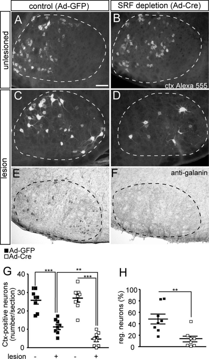
SRF depletion reduces facial nerve regeneration. A–D, The facial nerve of Srfflex1/flex1 animals was unilaterally lesioned. Seventeen days after axotomy, a fluorescently labeled tracer (Ctx Alexa 555) was injected and allowed to retrogradely label facial motoneurons for 4 d. After a total of 21 d, tracer-positive facial motoneurons were quantified in unlesioned (A, B) and lesioned (C, D) facial nuclei. On the unlesioned side, numbers of facial motoneurons in Ad-GFP- (A) and Ad-Cre (B)-injected animals were similar. On the lesioned side of control animals injected with Ad-GFP, robust axon regeneration was observed (C). In contrast, SRF ablation upon Ad-Cre injection decreased numbers of Ctx-positive neurons on the lesioned side (D). E, F, Galanin was found on the lesioned side of Ad-GFP (E), but only sparsely in Ad-Cre (F)-infected animals. G, Quantification of average Ctx-positive neuron number/section for the unlesioned and lesioned facial nucleus. The number of tracer-positive neurons on the lesioned side was ∼3-fold reduced upon Ad-Cre injection compared with Ad-GFP. H, The percentage of regenerating neurons is depicted by the ratio of neurons present on lesioned and control side. In Ad-GFP-injected animals, 50% of neurons regenerated. In contrast, only 15% of SRF-deleted neurons incorporated the tracer. Individual squares reflect independent animals analyzed. Facial nuclei are delineated by dashed lines. Scale bars in A–F, 100 μm.