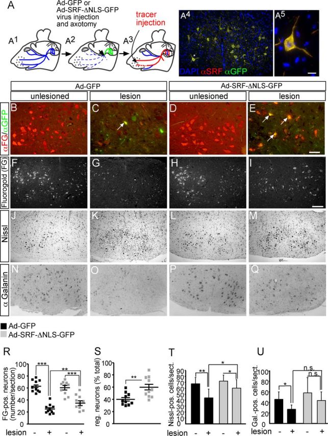
Figure 4.

Cytoplasmic SRF facilitates facial nerve regeneration in vivo. A, A1, The facial nerve is outlined in blue. A2, Ad-GFP or Ad-SRF-ΔNLS-GFP virus (green) was injected. The injection and axotomy position is depicted by an arrow. A3, Tracer (FG, red) is injected in the whisker pad and retrogradely transported to cell bodies of regeneration or sprout forming neurons. A4, Lesioned facial nucleus of an Ad-SRF-ΔNLS-GFP-injected animal. Overexpression of SRF-ΔNLS-GFP resulted in cytoplasmic SRF localization. A5, Higher magnification of neuron labeled by asterisk in A4. B–E, Inspection of axon regeneration of GFP expressing neurons only. The number of FG-positive neurons on the unlesioned side was comparable between both virus types (B, D). In Ad-GFP-infected animals, ∼30% of all neurons expressing GFP were also tracer positive (labeled with an arrow in C). In contrast, >70% of SRF-ΔNLS-GFP-expressing motoneurons were positive for the regeneration, indicating tracer (labeled with arrows in E). F–I, Visualization of the total population of FG-positive motoneurons in control (F, H) or lesioned (G, I) facial nuclei of GFP- (F, G) or SRF-ΔNLS-GFP (H, I)-expressing animals. SRF-ΔNLS-GFP enhanced the number of tracer-positive neurons in lesioned facial nucleus (I) compared with GFP (G). J–M, Nissl staining labels all facial motoneurons. In Ad-GFP-infected animals, the number of motoneurons was decreased upon lesion (J, K). In contrast, SRF-ΔNLS-GFP prevented motoneuron loss upon facial nerve injury (L, M). N–Q, In Ad-GFP-expressing facial nuclei, galanin-positive neurons were downregulated upon lesion (N, O). In contrast, more galanin-positive neurons were present upon lesion in animals expressing SRF-ΔNLS-GFP (P, Q). R, Quantification of axonal regeneration counting all FG tracer-positive cells (each square depicts one animal). The average number of FG-positive neurons/section upon lesion is increased by SRF-ΔNLS-GFP. S, Percentage of tracer-positive neurons in the total motoneuron population depicted by the ratio of neurons present on the control and lesioned side. T, U, SRF-ΔNLS-GFP increased the number of Nissl- (T) and galanin-positive (U) neurons upon facial nerve injury. Scale bars: A4, F–Q, 100 μm; B–E, 10 μm; A5, = 5 μm.
