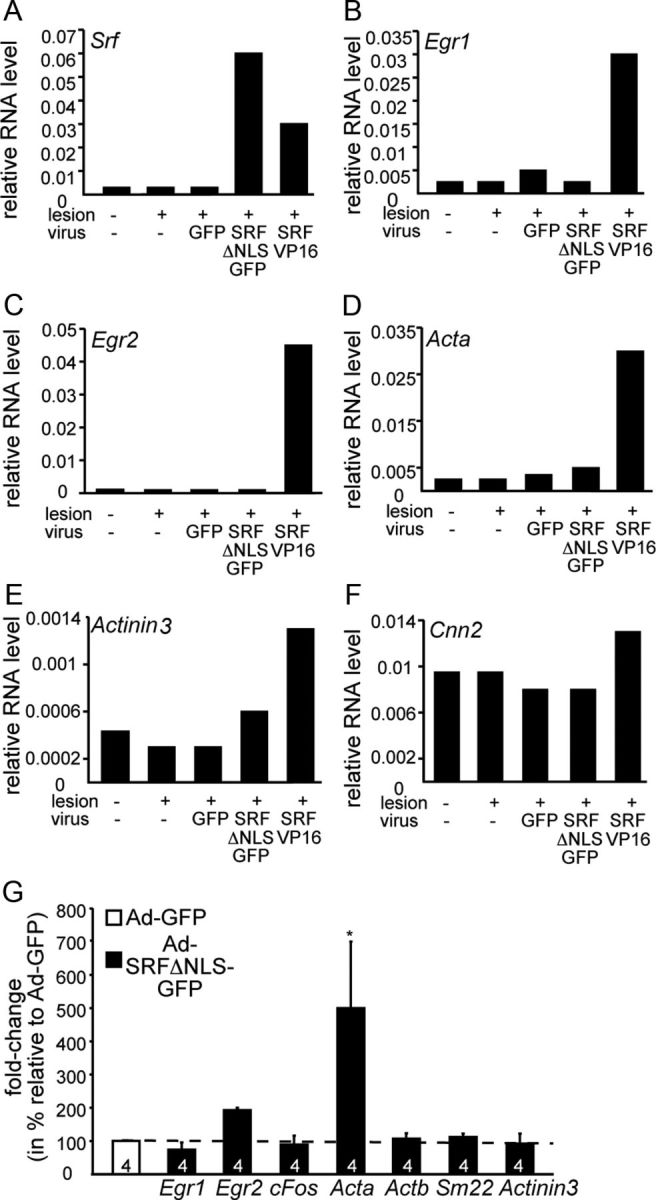
Figure 5.

Impact of cytoplasmic SRF on gene expression. A–F, Facial motoneurons were-infected with Ad-GFP, Ad-SRF-ΔNLS-GFP, or Ad-SRF-VP16. Three days later, cDNA of the unlesioned and lesioned sides was subjected to qPCR; primer pairs are indicated. SRF-VP16 induced mRNA levels of Egr1 (B), Egr2 (C), Acta (D), Actinin 3 (E), and Cnn2 (F). In contrast, SRF-ΔNLS-GFP had almost no effect on mRNA abundance of genes depicted. G, Cortical neurons were infected with Ad-GFP or Ad-SRF-ΔNLS-GFP. Three days later, mRNAs were isolated and cDNA was subjected to qPCR; primer pairs are indicated. SRF-ΔNLS-GFP only affected mRNA abundance of Egr2 and Acta and had almost no effect on mRNA abundance of all other genes depicted.
