Figure 2.

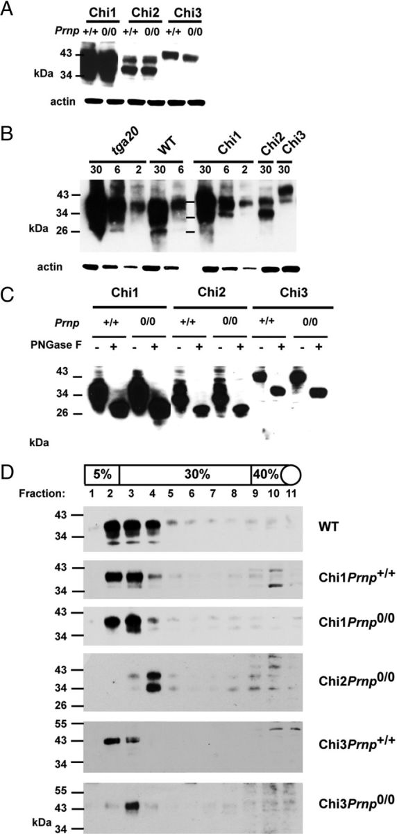
Expression of Tg proteins. A, The expression levels of each chimeric protein are independent of the Prnp status (Prnp+/+ or Prnp0/0). Thirty micrograms of protein from crude brains homogenate were loaded. B, Expression of chimeric protein in brain compared with that of PrP in WT and tga20 mice. Chi3 expression is lower than that of WT PrP. Amounts of protein (in micrograms) loaded for each line are indicated on the top of the gel. C, The glycosylation pattern of full-length Chi proteins is similar in Chi Tg mice expressed on mouse Prnp+/+and Prnp0/0 background. Thirty micrograms of protein from total brain homogenates are subjected (+) or not (−) to PNGase F treatment. D, Analyses of density gradient of detergent-resistant membranes from brains of WT, Chi1, Chi2, and Chi3 mice (on Prnp+/+ and Prnp0/0 background). Western blots were performed with mAb SAF32 (B, D; ChiPrnp0/0 Tg mice) or mAb 3F4 (A, C, D; ChiPrnp+/+ Tg mice). Molecular size markers (in kilodaltons) are indicated on the left.
