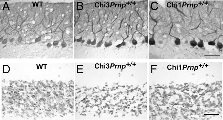
Figure 6.
Calcium-binding protein immunoperoxidase staining of the Purkinje cells in the cerebellum of 8-month-old WT (A) and Chi3Prnp+/+ (B) mice and 11-month-old Chi1Prnp+/+ mice (C). NeuN immunohistochemical staining of the GCs in the median cerebellar vermis of 8-month-old WT (D) and Chi3Prnp+/+ (E) mice and 11-month-old Chi1Prnp+/+ mice (F). Scale bars, 50 μm.

