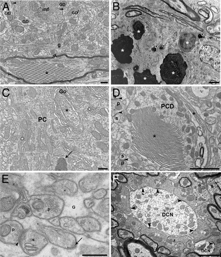
Figure 8.
Ultrastructure analysis. A, B, Ultrastructure of the internal granular layer of Chi3 mice. A, A mossy fiber terminal (mf) makes asymmetric synapses (arrowheads) with GC dendrites (GD) and is surrounded by abnormal wraps of glial profiles (g). A myelinated PC-like axon contains an abnormal crystalloid stacking of tubular profiles (*). B, Steps of GC degeneration from shrunken cell body with cytoplasmic electron-opaque profiles (arrows) to overall electron-opacity of the neurons (*). See the normal-like GC on the right side. C–F, Ultrastructure of abnormal profiles in PC somata and dendrites of Chi3 mice. C, Autophagic-like phagophore (*) forming from the trans saccules of a Golgi (Go) apparatus dictyosome wrapping in a PC body. Note also the abnormal stacking of endoplasmic reticulum (white asterisks) and a dense body (arrow). D, A main PC dendrite (PCD) displays abnormal stacking of endoplasmic reticulum (*). Arrowheads indicate asymmetric synapses between presynaptic parallel fiber boutons (p) and postsynaptic PC dendritic spines (s). E, A PC dendritic spine (white s) is completely surrounded by glia (G) and displays a postsynaptic density (arrow) free of a presynaptic partner. Another PC spine (s) is innervated (arrowheads) by a presynaptic parallel fiber bouton (p). F, PC axon terminals (Pa) make symmetric synapses (arrowheads) on dendrites of a deep nuclear neuron (DCN). Scale bars: A, E, 500 nm; B, C, D, F, 2 μm.

