Figure 3.
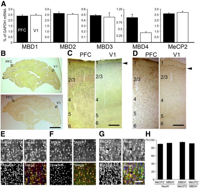
Expression patterns of methyl-binding proteins in macaque neocortex. A, The expression levels of MBD1, MBD2, MBD3, MBD4, and MeCP2 in PFC (black bars) and V1 (white bars) were examined by real-time RT-PCR. The amount of each mRNA was normalized as the relative ratio to that of the internal standard, GAPDH mRNA. MBD4 expression level in PFC was higher than that in V1, whereas all four other genes showed no significant difference in expression level between PFC and V1. Error bars indicate SD. B, Parasagittal sections of the adult macaque brains were stained for MBD4 by both ISH (top) and IHC (bottom). Right, Higher magnification photomicrographs of the boxed regions of PFC and V1 (C, ISH; D, IHC). Scale bars: B, 10 mm; C, D, 400 μm. E, MBD4-positive cells express the neuronal marker NeuN. Hoechst dye was used for nuclear staining. All three markers are merged in the right bottom part. F, MBD4 and MeCP2-positive cells highly overlapped. G, MeCP2-positive cells also express the neuronal marker NeuN. Scale bar, 50 μm. H, Quantification of MeCP2- and MBD4-positive cells in NeuN-positive cells. The total proportion of MBD4-positive cells among MeCP2-positive cells, and vice versa, is shown. Three 200 × 200 μm2 windows within layer II in PFC (from three monkeys) were used for counting. Error bars indicate SD.

