Figure 5.
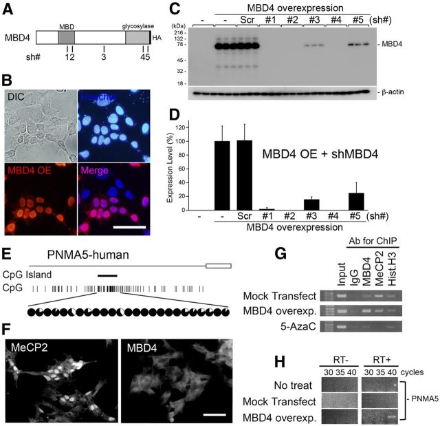
Overexpression of MBD4 induces the expression of PNMA5 mRNA in human neuroblastoma cell culture. A, Schematic illustration of the full MBD4 protein with an HA-tag in the C terminus, and the positions of the 21 bases of the sh #1–5 vectors used for MBD4 knockdown. B, Overexpression (OE) of MBD4-HA in SH-SY5Y cells. The overexpressed proteins were stained with the anti-HA antibody (red). The localization of MBD4-HA in the nuclei was determined by costaining with Hoechst (blue). C, D, Quantification of the knockdown effects of cotransfection of the U6-shMBD4 (#1–5) vectors with MBD4-HA in SH-SY5Y. Sh-MBD4#4 (with the highest efficiency, 99% knockdown effect) was used in subsequent in vitro and in vivo experiments. Error bars indicate SD. E, The human PNMA5 promoter has a CpG island at a position similar to that in the macaque monkey (Fig. 2A, PNMA5), and this island is also hypermethylated in SH-SY5Y human neuroblastoma cells. F, SH-SY5Y cells express MeCP2 and not MBD4. G, MeCP2 is bound to the CpG islands in the human PNMA5 promoter in control conditions (Mock transfect/MeCP2:IP), whereas blocking methylation by 5-AzaC diminishes this binding (5-AzaC/MeCP2:IP). Overexpressed MBD4 may bind at the same site because MeCP2 binding appears to have been reduced (MBD4 overexp/MBD4 and MeCP2:IP), but this result remains to be confirmed, as the transfection efficacy to the cells was not 100%. H, The overexpression of MBD4 induced PNMA5 expression in SH-SY5Y cells.

