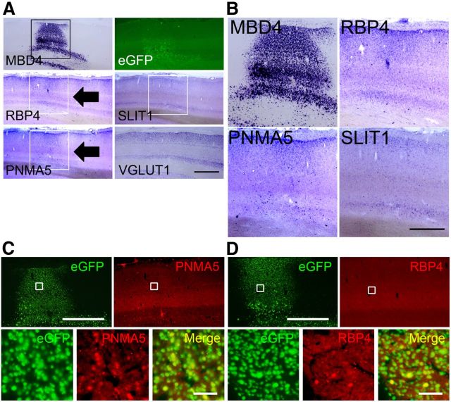
Figure 7.
Expressions of AA-selective RBP4, PNMA5, and SLIT1 in the AAV-MBD4-injected monkey V1. A, Expressions of AA-selective RBP4, PNMA5, and SLIT1 in sections of V1 in AAV-MBD4-injected monkey. ISH signals of the ectopic expression of both PNMA5 and RBP4 observed in the MBD4-injected sites in V1 (black arrows). The white boxes for MBD4, RBP4, and PNMA5 indicate the injection sites within these areas showing ectopic expressions of these genes. The photomicrographs of eGFP, SLIT1 (indicated by white boxes), and VGLUT1 expressions around the injection site are also shown. Scale bar, 2 mm. B, Higher magnifications of these boxed areas, demonstrating the ectopic expressions of MBD4, RBP4, and PNMA5, but not that of SLIT1. Scale bar, 1 mm. C, D, Double-labeling fluorescence ISH of AA-selective genes (red) and transgened marker eGFP (green). Although the expression levels of PNMA5 and RBP4 varied, we were able to confirm that the ectopic expressions of PNMA5 (C) and RBP4 (D) in V1 colocalized well with eGFP expression, which demonstrated that the expressions of these genes were affected in the same cells as those with the genetic manipulation. Note that, in the loss of function experiments in PFC, the MBD4 and PNMA5 signal intensities were too low to be detected by our double-ISH protocol (data not shown), by which a signal is more difficult to be detected than by single-labeling ISH (Fig. 6C,D). Scale bars: (for C, D), top, 2 mm; bottom, 50 μm.

